
New version (June, 2020)
This is a new version of X-Ray Server's diffraction program launched in
June-2020. Compared to the previous version, it provides the following
benefits and features:
- Up to 10x faster calculations due to the reduction of the scattering
matrix size. The original GID_sl was designed for Grazing
Incidence X-ray diffraction (GID) where both incident and diffracted
waves make small angles to the crystal surface and experience specular
reflection. In this case the scattering matrix is 4x4,
which corresponds to accounting for not only incident and diffracted waves,
but also their specularly reflected counterparts. However, in the vast
majority of calculations performed at X-Ray Server GID_sl
is used for usual diffraction without the GID effects. In these cases the
specularly reflected waves can be neglected and the scattering matrix can
be reduced to the 2x2 size. Similarly, in the extremely asymmetric cases,
whether grazing incidence or grazing exit, the scattering matrix can be
made 3x3. The new version of GID_sl introduces the scattering
matrix reduction parameter, which may have tree values: No (the old 4x4
behavior), Prescan and Fly. When the parameter is set to "Prescan",
GID_sl examines all Bragg curve points for possible reduction
and then applies the maximum found matrix size to all points. This option
ensures there is no matrix size switch during the scan. With the "Fly"
option, the matrix size is chosen individually for each point of the scan,
which is the fastest. If all Bragg curve points are calculated with the
2x2 matrix the calculation time is reduced by approximately 10x compared
to the 4x4 case.
- Option xhphase to specify phases for the Xh (Chi-h) parameters
of the layers. This is mainly meant for very thin layers which may grow
on substrates as e.g. either ABAB... or BABA... Note that xhphase
differs from xhdf which is a phase shift between Xrh and Xih in
non-centrosymmetric crystals. The phases are specified in the multiples
of π and can be in the range [-2, 2]. Example:
xhphase=1.
- Option to calculate X-ray Standing Waves (XSW)
in crystals. One can calculate either the XSW at certain depth as a
function of scan parameter (angle or energy) or a 2D-pattern as a
function of scan parameter and the depth inside the crystal. Two modes
are available: either the phase in π units of the probe point
location with respect to the Bragg planes (0=in planes, 1=between the
planes) or let the phase change with depth when the phase input is left
blank or specified as "none".
- New calculation engine based on the
ZGESV (LAPACK) and
CPOLY (ACM)
open-source routines.
This page is a CGI interface to my program GID_sl simulating dynamical
x-ray diffraction from strained crystals, multilayers, and superlattices. The
name 'GID_sl' comes from the original aim of this program to simulate
grazing incidence x-ray diffraction from multilayers and superlattices [1-3]. Later on, GID_sl was extended to extremely
asymmetric geometries and usual Bragg-case [4]. At
present this program is applicable to any coplanar
and non-coplanar
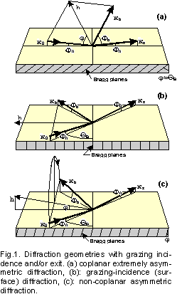
Bragg-case geometry with diffraction scans round arbitrary axes. Some typical
examples of grazing-incidence geometries where the GID_sl can be used
are shown on Fig.1.
The program can simulate the following profiles of structure parameters and
defects in multilayers:
- Normal lattice strains, da(z)/a;
- Crystal polarizabilities x0(z), xh(z),
[chi0, chih] including the Debye-Waller
profiles wh(z), w0(z);
- Interface roughness sigma(z) in multilayers.
The effects of lateral strains (relaxed multilayers, misfit dislocations,
etc) or crystal curvature are NOT simulated by this code. GID_sl is
not aimed either for the truncation rod scattering (for very big deviations
from Bragg peaks).
The program implements a "discrete" algorithm, i.e. the surface
layer of crystal is subdivided onto "perfect" sublayers, each one
possessing its own x0, xh, normal strain da/a, thickness
t, and the rms height sigma of interface roughness. After solving
the dynamical diffraction problem for each of the layers, the reflection from
the whole system is calculated with the help of the (2x2) recursive matrix
algorithm, RMA [6], which is a development of our earlier
(4x4) transfer matrix approach, TMA [1-5]. While the TMA
was an extension to the Abeles matrix solution [7] for
x-ray specular reflection by multilayers, and the RMA can be viewed as a
generalization of Parratt's and Bartels' recursive methods [8,9] for the specular reflection and non-grazing Bragg
diffraction by multilayers respectively. The RMA works about 2 times
slower than the TMA, but it is free of some principal divergencies which
might happen with the TMA. In fact, GID_sl dynamically selects between
the TMA and RMA depending on the conditions of calculations.
Acknowledgments: The idea of this X-ray Web Server appeared under
the influence of excellent
x-ray server by Eric
Gullikson at the LBL. The solution to the plots outputs was
prompted by William Lavender.
Some ideas on making GID_sl user-friendly were suggested by Pavel Petrashen.
I am grateful to all of my co-authors listed in the References below and to many
others for the collaboration and helpful discussions resulted in the development
of the TMA and RMA methods. Finally, since the server was launched, I have received
a number of valuable comments and suggestions from many users. This response cannot
be overestimated because it helps to improve the algorithms, extend the services
and avoid possible bugs. Among many othes I am especialy grateful to
Prof. Dr. Marius Grundmann
for the communication helped to extend the calculations to larger strains and
for comparing GID_sl with available commercial software [10].
References
- S.A.Stepanov,
"Method
of transfer matrices and dynamical thick-crystal approximation in surface x-ray
diffraction by multilayer structures", Crystallogr. Reports,
39 (1994) 182-187.
- S.A.Stepanov, U.Pietsch and G.T.Baumbach,
"A matrix approach to x-ray grazing incidence
diffraction in multilayers", Z. Physik B, 96 (1995)
341-347.
- S.A.Stepanov and R.Koehler,
"Real-structure
effects in the dynamical theory of grazing incidence x-ray diffraction",
J. Appl. Phys., 76 (1994) 7809-7815.
- S.A.Stepanov and R.Koehler,
"A
dynamical theory of extremely asymmetric x-ray diffraction taking account of
normal lattice strain" , J. Phys. D: Applied Physics, 27
(1994) 1923-1928.
- S.A.Stepanov, E.A.Kondrashkina, M.Schmidbauer, R.Koehler, J.-U.Pfeiffer,
T.Jach, and A.Yu.Souvorov,
"Diffuse
scattering from interface roughness in grazing incidence x-ray diffraction",
Phys. Rev. B, 54 (1996) 8150-8162.
- S.A.Stepanov, E.A.Kondrashkina, R.Koehler, D.V.Novikov, G.Materlik, and S.M.Durbin,
"Dynamical
x-ray diffraction of multilayers and superlattices: Recursion matrix extension to
grazing angles", Phys. Rev. B, 57, (1998) 4829-4841.
- F.Abeles, Ann. Phys. (Paris), 3 (1948) 504; 5 (1950) 596.
- L.G.Parratt, "Surface studies of solids by total reflection of
x-rays", Phys. Rev., 95 (1954) 359-369.
- W.J.Bartels, J.Hornstra and D.J.W. Lobeek,
"X-ray diffraction of multilayers and superlattices",
Acta Cryst. A, 42 (1986) 539-545.
- M.Grundmann and A.Krost,
"Atomic structure based simulation of X-ray scattering from strained
superlattices", Phys. Stat. Sol. (b), 218 (2000) 417-423.
This short guide provides some explanations on the GID_sl
data input and outlines the restrictions of this Web interface.
The GID_sl program is executed on a single PC, which runs a Web server
under Windows operating system. Since this PC is shared by all X-ray Server users,
there is a mechanism to restrict the maximum number of tasks processed at a time.
If you unlikely hit that restriction, please try back later. Also, please,
avoid overloading the server by submitting multiple tasks at the same time.
To obtain the results from GID_sl one needs to fill out the input form
and click on the SUBMIT button. If the input is correct, the results will be
presented as a figure and a reference to downloadable ZIP file containing the
data and the listing of calculation parameters. Otherwise, an error message
will be returned.
The specification of x-rays should not cause any problems. If mixed
polarization is chosen, the results for σ- and π- polarizations are
added with the weight contributions 1/(1+|cos(2*QB)|) and |cos(2*QB)|/
(1+|cos(2*QB)|), respectively. The weights are interchanged for the special
case of scan which simulates GID takeoff spectra measured with
position-sensitive detector (PSD). In these measurements the monochromator is
turned by 90 degr. and it collimates the incidence angle instead of beam
divergence in the Bragg plane.
The crystal substrate material is specified by the "crystal code"
referring to the X0h database which is used for automatic
calculation of the scattering and absorption factors x0 and xh (chi 0
and chi h) respectively. See the X0h
page for more information on the scattering factors calculations. Here is
the
list of crystals currently available in the X0h database.
Then, "Reflection" stands for the Miller indices of the Bragg
reflection; "Sigma" is the rms height of substrate roughness
expressed in Angstrom; "W0" and "Wh" are the Debye-Waller
like corrections to the values provided by the X0h database for
x0 and xh respectively. Finally, "da/a" is the correction to the
Bragg planes spacing (the X0h data are used as a reference
value).
The geometry of Bragg diffraction can be specified by 9 different ways.
Basically one has to distinguish between the coplanar diffraction cases
(those where the incident wave vector k0, the reciprocal
lattice vector h, and the diffracted wave vector kh
lay in the same plane perpendicular to the surface -- Fig.1a) and the
non-coplanar cases (Fig.1b,c) where the above condition is not satisfied,
i.e. the scattering plane defined by k0 and h is
not perpendicular to the surface. The coplanar geometries are more commonly
used in the x-ray experiments since they are simpler to implement. On the
other hand, the non-coplanar geometries provide a greater flexibility in
choosing the x-ray incidence and exit angles as well as in scanning around
the Bragg peaks.
If one knows the orientation of crystal surface, then the coplanar diffraction
cases (Fig.1a) can be specified as either grazing incidence, or grazing exit,
or the symmetric Bragg case. In the latter case the Bragg planes must be
chosen parallel to the surface.
Four simplified inputs are provided for coplanar diffraction geometries.
Those do not require a knowledge of crystal surface orientation, but instead
they expect one of the four parameters: either the incidence angle of
k0, or the exit angle of kh, or
disorientation angle of the Bragg planes, or the asymmetry factor
beta=g0/gh. Use the SIMPLIFIED TEMPLATES below to
access the new interfaces.
The non-coplanar cases (Fig.1b,c) always require the specification of crystal
surface orientation and an additional parameter -- either the incidence or
exit angle for the exact kinematical Bragg position (the actual
position of the Bragg peak may deviate from the kinematical value due to
refraction). The specification of additional angle is not required for
symmetric non-coplanar cases (Fig.1c). The grazing incidence diffraction
geometry (Fig.1b) as a particular case of non-coplanar geometries, always
requires specifying the incidence or exit angle of x-rays. A common mistake
is specifying GID as a symmetric Bragg case. Then, as one can see on Fig.1b,
the incidence and exit angle will be taken zero. This happens because GID is
a symmetric Laue case, not the Bragg one, and the wave yielding the crystal
in GID is a specular component of diffracted wave, not the real one.
For more details on the specification of coplanar and non-coplanar
geometries to GID_sl click here.
Here are some examples for GaAs crystal with the (001) surface plane specified:
- The (004) reflection can only be specified as a symmetric Bragg
case (there is no choice).
- Reflection (113) can be specified as an extremely asymmetric
coplanar case with grazing incidence. For E=8.5 keV this gives the
incidence angle of 0.09 degr.
- If a higher incidence angle is required, one can proceed to
non-coplanar case and select the incidence angle equal to e.g. 1.00 degree.
- Finally, the same (113) plane can be brought into the symmetric
non-coplanar geometry (in this case the incidence and exit angles will be
equal to 22.77 degr.)
The crystal surface orientation (if needed) is specified via the indices of
external surface normal, the angle and the direction of maximum miscut.
Please, do not specify non-integer indices if the miscut direction does not
coincide with some crystallographic axis: use an integer approximation
instead. Example: enter (111 322 4) instead of (1.11 3.22 0.04).
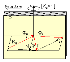 The scan axis for coplanar cases is usually a vector perpendicular the
scattering plane and parallel to the surface. Such a vector can be presented
as [k0 x h] or [k0 x N_surface] -- Fig.2. For the grazing incidence
diffraction (Fig.1b) the scan vector is usually N_surface (the crystal is
rotated round the surface normal). GID_sl can also simulate the
special case of GID where the incidence beam is not collimated in the Bragg
plane and the sample is not scanned. Instead of scanning, the diffraction
curve is recorded by PSD. For other non-coplanar cases there is a choice of
several "natural" scan axes, or an arbitrary axis can be specified (please,
use integer indices).
The scan axis for coplanar cases is usually a vector perpendicular the
scattering plane and parallel to the surface. Such a vector can be presented
as [k0 x h] or [k0 x N_surface] -- Fig.2. For the grazing incidence
diffraction (Fig.1b) the scan vector is usually N_surface (the crystal is
rotated round the surface normal). GID_sl can also simulate the
special case of GID where the incidence beam is not collimated in the Bragg
plane and the sample is not scanned. Instead of scanning, the diffraction
curve is recorded by PSD. For other non-coplanar cases there is a choice of
several "natural" scan axes, or an arbitrary axis can be specified (please,
use integer indices).
The scan range is that around the Bragg peak along chosen scan axis. The
GID_sl allows plotting the diffraction curve depending on the scan
angle, the incidence angle, or the exit angle. However, in all of the cases
one has to specify the scan range as the deviations from the Bragg peak. It
is a frequent mistake when users request the plot depending on the incidence
angle of x-ray and then attempt to specify the incidence angles in the scan
range fields.
The alpha_max tells GID_sl at what Bragg deviations
alpha=((k0+h)^2-k0^2)/k0^2 it can stop accounting for diffraction in
layers and switch to an amorphous layer model. At large deviations
from the Bragg condition the diffracted wave intensity is very weak
and trying to account for this tiny effect may lead to loss of
precision in the calculations. It is recommended to leave this
parameter unchanged unless you experience some numerical errors.
The scattering matrix reduction parameter allows GID_sl to
reduce the scattering matrix size from 4x4 when either the incident
or the diffracted wave or both are not grazing. It may speed speedup
calculation up to 10 times and improve stability by not trying to account
for very weak effects along with the strong ones. This parameter may
have tree values:
- No which means always use 4x4 scatting matrix where all
specular waves are taken into account. This corresponds to the
behavior of the previous GID_sl version.
- Prescan instructs GID_sl to examine all Bragg
curve points for possible reduction and then apply the maximum found
matrix size to all points. This option ensures there is no matrix size
switch during the scan.
- Fly instructs GID_sl to choose the matrix size individually
for each point of the scan. The size may be chosen as 4x4, 3x3, and 2x2.
This is the fastest and the default.
Along with the Bragg curves, GID_sl can also optionally calculate
X-ray Standing Waves (XSW) in the layers (see an XSW
template below). It can calculate either the XSW curves as a function
of the scan angle or the maps with the depth inside the crystal as the
second coordinate. While most of the XSW parameters are probably
self-explanatory, the Location phase parameter is the location of
probe point with respect to the Bragg planes measured in π:
when phase=0 the probe is located in the planes and when phase=1 the
probe is located midway between the planes. When the phase is not
specified, it is taken varying with the depth coordinate z.
The specification of crystal surface layer profile is implemented with
a simple scripting language. A typical syntax is:
; comments are allowed in any line, but should
; not contain special symbols like '"*?$!@%
period=5
t=10 x0=(5e-4,7e-6) xh=(3e-4,5e-6) xhdf=1. w0=1, da/a=2e-4
t=10 code=GaAs x=0.3 code2=AlAs x2=0.7 da/a=auto sigma=2
t=10 code=InAs xhpase=1
t=10 wh=.5 da/a=2e-4 sigma=2
t=10 da/a=2e-4
t=10
end period
-- where:
Here is a practical example -- a profile for 20-period AlAs/GaAs superlattice
with 100 Angstroms of GaAs and 70 Angstroms of AlAs in each period; the structure
is covered by additional 200A of GaAs and, finally, there is some 20A amorphous
oxide layer on the surface:
; Oxide layer:
t=20. w0=0.7 wh=0.
; -- w0=0.7 because of reduced layer density
; -- wh=0 since the layer is amorphous

; Cap layer:
t=200.
; -- when code is not specified, then
; the substrate code (GaAs) is used

; Superlattice:
period=20
  t=100.
  t=70. code=AlAs da/a=2.775e-3
  ; The strain value is taken from Bocchi et al.,
  ; J.Cryst.Growth, 132 (1993) 427
end period
For the rest of parameters users are suggested to follow their common sense.
To ensure that the input was correct, please verify respective listing file
-- a file with the ".TBL" extension in the ZIPped archive
returned by GID_sl.
To simplify understanding the GID_sl interface, users are provided with
the templates listed below. All of the templates link to the same program and
provide the same functionality (except for the simplified templates which are
designed for coplanar diffraction only). The templates differ by preloaded data
to demonstrate some possible applications of the GID_sl.
When submitting the GID_sl task, it is possible to check the progress
watching option. The progress watching is obviously more comfortable, but it
might not work with some old Web browsers. Also, it is a bit slower because
of putting an additional load on the network and launching each 5 seconds a
status query program on my computer. Welcome to try both of the ways and
choose the most convenient for your needs.
Here is a tool to retrieve the results of finished jobs if you know the job ID.
Some possible uses of this tool are:
- You started a job with the progress watch option; the server returned
the job ID and began reporting the progress. However, you found that the
calculations would take too long. Then, you may break the connection and
retrieve the data later on with this tool. If the calculations are not
finished, the tool will resume the watch process.
- The data are accidentally deleted from your client computer and you want
another copy of them. In this case you should be aware that results are
usually stored on the server for about one day after respective job is
finished.
